Professor Kazem Fathie, M.D., F.A.C.S., F.I.C.S., Ph.D., KINGSLEY B. GRANT, M.D., and JOHN F. HESS, M.D.
With the exception of multiple myeloma, osteogenic sarcoma is the most common ma-lignant primary tumor of bone.(1) However, reports of its location in the vertebral column, particularly the cervical region, are extremely rare.(2,3) The purpose of this paper is to record a case of osteogenic sarcoma in the cervical vertebra of a 40-year-old Caucasian male.
CASE HISTORY
This 40-year-old white male complained of right-sided neck pain when first seen in February 1969. The patient related a history of spontaneous onset of cervical pain in 1965 which was relieved by chiropractic manipulations. In the spring of 1968, the pain recurred and at that time neither chiropractic treatment nor symptomatic care by a physician were ef- fective. X-rays of the cervical spine and skull in January 1969 were interpreted as normal with the incidental finding of cervical ribs. However, in retrospect, there was a small irregularity along the right lateral mass of the atlas. An orthopedist diagnosed “myofascial cervical strain” and after a trial of traction, a cervical cast was applied. The patient showed no improvement and on May 31, 1969, he was admitted to St. Luke’s Methodist Hospital with neurological findings of 7th and 12th nerve involvement on the right.

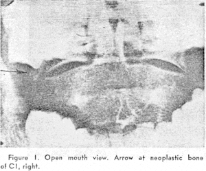
figure 1. Open mouth view. Arrow at neoplastic bone of C1, right
There was deviation of the tongue with numbness along the lateral portion, dysphagia, hoarseness, and slight dysarthria. He complained of nocturnal pains at the base of the skull, radiating into the right ear and right side of the head, along with severe pressure sensation in the back of the neck. Extensive neurosurgical evaluation, including right common carotid and cerebral angiography, brain scanning, electroencephalography, echoencephalography, complete myelography and air studies, produced findings entirely within normal limits. X-rays of the skull, cervical spine and mastoids were thought to be within normal limits (but in retrospect, an area of irregularity, along the right lateral mass of the atlas is noted, and is prominent on the open mouth views of the odontoid). (Figure 1) Laboratory examination showed: white blood cells 5,600; Hemoglobin 14.5; VDRL negative. A large, hard mass behind the mastoid process in the region of the stylomastoid foramen was palpable on the right side of the neck. This mass was painful and extended almost to the base of the skull. A biopsy from the region of the mass was negative for tumor. On June 27, 1969, the patient was discharged for office follow up.
OPERATION
The patient was readmitted in October 1969 for posterior auricular and occipital neurectomies because of his increasing pain and disability.

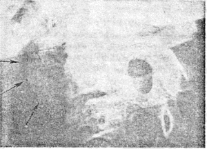
Preoperative films of the cervical spine, mastoid region and base of the skull re- vealed a large area of tumor calcification along the right border of the atlas and adjacent to the base of the skull. (Figure 2) Repeat biop- sy from the deep tissues of the neck revealed a highly anaplastic tumor of malignant osteoid formation in an extremely fibrocellular stroma (Figure 3), along with marked hyperchromasia and numerous bizarre mitotic fig- ures (Figure 4). A final diagnosis of osteogenic sarcoma was made.
POST-OPERATIVE COURSE
The patient was treated at the Mayo Clinic to maximum tolerance with radiation and chemotherapy. He pursued a steadily deteriorating course and at terminal admission to St. Luke’s Methodist Hospital was unable to fully open his mouth because of mastoicl and maxillary infiltration by the tumor. He expired on September 1, 1970. At postmortem both lungs were the site of massive fibrous, osteoid and frank bone tumor metastases (Figure 5), with extensive secondary bronchopneumonia. The liver, kidneys and lymph nodes also contained metastases. Cosmetic considerations precluded adequate exploration of the primary mass in the right neck and face but sections of the strap muscles showed diffuse infiltrating tumor. No intracranial lesions were present.
DISUSSION
In their review of 430 cases of osteogenic sarcoma, Coventry and Dahlin(1) analyzed extensively the histologic and biologic features of these tumors. That review, including the discussants’ remarks, and the material from more recent texts(4, 5) are sufficiently cogent and applicable as not to warrant further detailed commentaries in this report.
SUMMARY
A case report of osteogenic sarcoma arising in a cervical vertebra of a 40-year-old white male is presented. The authors stress the rarity of this location for what is the second most common malignancy of bone.
REFERENCES
1. Coventry, M. B., and Dahlin, D. C.: Osteogenic sarcoma -critical analysis of 430 cases. J. Bone Joint Surg., 39-A:741-758,1957. 2. Cohen, D. M., et at: Apparently solitary tumors of vertebral column. Mayo Clin. Proc., 39:509-528,1964. 3. Hastings, D. E., et at: Neoplasms of atlas and axis. Canad. J. Sur,11:290-296,1968. 4. Ackerman, L. V., and Spjut, J. J.: Tumors of bone and cartilages A.F.I.P. Atlas Tumor Patho. Section II: Fascicle 4, 84-87,1962.
