Professor Kazem Fathie, M.D., F.A.C.S., F.I.C.S., Ph.D.
CAROTID ARTERY OCCLUSION is caused by atherosclerosis, arteriosclerosis and atheroma, and is compounded by the extension of cholesterol and calcium deposits into the branches of the common carotid artery, specifically the internal carotid artery. It has been known for some time that the end result of the cerebrovascular accident is rather massive. Devastating neurological symptoms appear such as paralysis of the upper and lower extremities or face, speech difficulties, visual blindness and/or aphasia. Transient episodes of clizziness, blackout spells and light headedness have been observed in many patients with partial occlusion of the internal carotid artery. Lowering of the blood pressure in the patient with partial occlusion of the carotid artery has been one of the prime factors causing permanent or irreversible neurological deficit. The surgical approach to the removal of the atheroma, called an endarterectomy, has not been consistently successful because long standing closure of the internal and common carotid artery presents a surgical hazard in resulting brain damage. Short term closure of the carotid artery has not allowed sufficient time for the surgeon to remove the atheroma completely from the wall of the arteries. Application of conventional shunting has been basically ineffective for correct perfusion of the arteries and brain.
It has been because of this important time consideration that we have developed a device called FATHIE SHUNT. The unit is a three- way shunt (Figure 1) made of medical-grade silicone rubber with imbedded wire reinforcement throughout to eliminate kinking and occlusion of the lumens. The shunt has been designed to fit the anatomical configuration of the internal, external and common carotid artery and has an inflation balloon at the end of each tube to occlude the space between the outside of the shunt branches and the respective arteries. Each balloon has its own small diameter inflation tube for individual control. When the shunt is applied and inflation
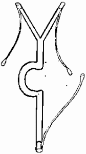
Figure 1. Diagram of Fathie Shunt
carried out, the surgeon is able to remove the temporary ligation of the internal, external and common carotid arteries allowing the blood to reach the brain in the specific amount previously received. The surgeon can take his time and make an incision to remove the endarterectomy plaque with the utmost accuracy and completeness. Initially this shunt was made in the shape of the artery but was later modified to allow the surgeon more room to work. This was accomplished by offsetting the :main section of the shunt to stay clear of the trunk of the vessel. Later, another modification distinguished between the right Fathie Shunt and the left Fathie Shunt, one for each side. Different size shunts have been made in both the right and left shapes to accommodate the various size arteries. Upon completion of the surgery, temporary ligation of the arteries is again applied, the Fathie Shunt removed, and the artery closed. During the preliminary stage of development, the shunt was initially used in the laboratory to determine the feasibility of clinical utilization. Once proven in the laboratory, clinical application of the device was made successfully in the operating room with minimal operative complications and good postoperative recovery. The application of this shunt is valuable when dealing primarily with partial occlusion of the carotid artery, and occasionally in the complete occlusion of the internal carotid artery with retrograde bleeding from behind the occlusion. In cases of internal carotid occlusion when there is no retrograde hemorrhage from behind the occlusion, there would be no need to apply the Fathie Shunt.
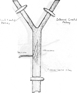
Figure 2. Incision in carotid artery showing location and outline of atheroma
SURGICAL TECHNIQUE
Prior to starting the surgery, all sizes of Fathie Shunts should be properly cleaned and sterilized. All inflation balloons should be checked for leakage using sterile, normal saline under aseptic conditions. This is necessary because of the delicate nature of the balloon construction which can be damaged by sharp instruments or rough handling. After opening the neck in the usual manner and exposing the internal carotid artery and external carotid artery, place an umbilical band under each artery and free each artery of periarterial tissues as well as surrounding neighboring tissues. Care should be taken to expose the external and internal arteries as completely and as high as possible and the common carotid artery as low and completely as possible. The size of the artery can then be observed and the proper size of Fathie Shunt selected for the shunting. The common carotid, external carotid and internal carotid arteries are occluded by application of the Bull- dog clamp. An incision approximately 1 1/2 cms in length is made over the anterior border of the common carotid artery (Figure 2). If there is any atheroma observed at the center portion of the artery, it is removed and the rest of the atherosclerosis left alone. The common carotid branch of the shunt is then placed in the common carotid artery (Figure 3) and inflation of the balloon carried out sufficiently to stop leakage around the shunt tube. Care is taken not to expand or stretch the artery to an abnormal size during the inflation. Remove the Bulldog clamp on the common carotid artery to allow blood to pass through the shunt and exit from the external and internal branches of the shunt.
[one_half]
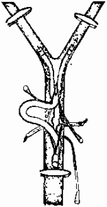
Figure 3. Insertion of Fathie Shunt
[/one_half]
[one_half_last]
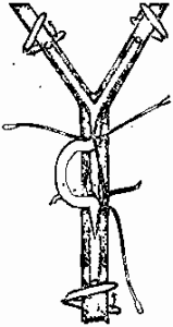
Figure 4. Fathie Shunt in position position showing inflation of balloons removal of lamps
[/one_half_last]
[one_half]
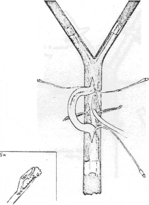
Figure 5. Fathie Shunt diverting the blood around the surgical field allowing removal of atheroma.
[/one_half]
[one_half_last]
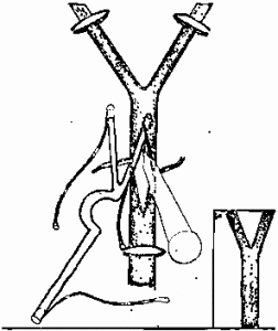
Figure 6. Removal of the shunt with ligation clamps in place. Irrigation utilized. Incision sutured closed.
[/one_half_last]
two branches are then occluded temporarily by Bulldog clamps or by placing a clamp in the lower portion of the common carotid artery of the shunt. Then the internal carotid artery portion of the shunt is inserted into the internal carotid artery making sure that no air is trapped in between the tube and the arterial wall by flushing the blood into the tubes and washing the arteries. The inflation is carried out again the same way (Figure 4) until the artery is closed; at this time, the internal carotid artery branch could be opened by removal of the clamp and the blood will rush from the common carotid artery into the internal carotid artery. The same procedure can now be performed on the third branch of the shunt into the external carotid artery. On some occasions, this branch can be left without any usage; however, we prefer to place the third branch into the external carotid artery, inflate the balloon with fluid, open the exter- nal carotid artery, and have the circulation established in all three routes. It should be mentioned that the left Fathie Shunt should be used for the left carotid artery and the right side for the right carotid artery in order to have the arch of the shunt always facing outside and away from the surgical field. It is also preferred that no air be injected into the inflating balloons and at all times the inflation be done by injection of saline solution or sterile water. In case the balloon becomes defective during surgery, this precautionary measure would ensure that no air would enter the arterial system to cause a cerebral embolism. The surgeon can now remove the atheroma from the wall of the artery accurately and ex- tensively without the pressure of time (Figure 5). After removing the atheroma, a heparin solution is applied to the arterial region and the area is irrigated with sterile, normal saline. The incision can now be closed to approximately 1 cm and Bulldog clamps again placed above the inflation balloons in each of the three branches. The Fathie Shunt is then removed from each vessel branch (Figure 6). The common carotid artery is unclamped first and irrigated with blood to eliminate air; then, one at a time, the clamps on the external and internal carotid arteries are released. During this procedure, Surgicell is utilized on top of the artery, and the incision is closed layer by layer in the conventional manner. It should be noted that the periods of time required for ligation of the arteries to insert the shunt device and to subsequently remove the shunt are both approximately two minutes in duration.
[one_half]
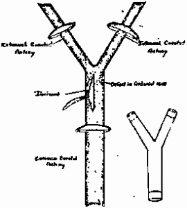
Figure 7. Preparation for insertion of permanent shunt
[/one_half]
[one_half_last]
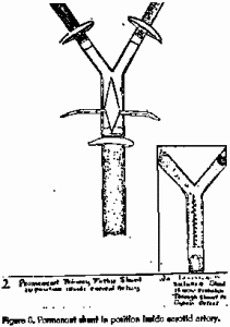
Figure 8. Permanent shunt in position inside carotid artery
[/one_half_last]
The two periods of occlusion have not caused any observed neurological deficit. We are also working on a further development for utilization in cases of endarterectomy when a defect is present in the wall of the common carotid arteries caused by atheroma and compression. The Shunt (Figure 7), a modification of the Fathie Shunt described above, would be placed in the artery and left permanently in order to prevent hemorrhaging or stricture of the mother vessels (Figure 8).
CONCLUSION
Further clinical investigation will be required to perfect this procedure so that, in the future, it can be performed less hazardously with the end result being quite valuable in the immediate recovery of stroke patients. It should be mentioned that, in the case of embolism, thrombosis, and the immediate sudden onset attack of occlusion, the previous method of endarterectomy and removal of clot would be as valuable as before.
SOURCE
The silicone rubber shunts described in this paper are manufactured by Heyer-Schulte Corporation, 5377 Overpass Road, Santa Barbara, California 93105. While this article is being published, the Heyer-Schulte Corporation has advised that seven centers in the United States and one in France are using the shunt. No study on its use in these locations has been completed.
